Education 645 High School Biology

Animal Cell Diagrams Labeled Printable 101 Diagrams
A labeled diagram of an animal cell, and a glossary of animal cell terms. Learn about the different parts of a cell. Label the Animal Cell. Label the Animal Cell Printout.. Plant and Animal Cells Venn Diagram: Use the Venn diagram below to list characteristics of plant and animal cells. Other Links:

Label the Parts of the Plant and Animal Cell Biology LibreTexts
Eukaryotic cells,one of the two major types of cells, have a nucleus. A nucleus is a large structure that controls the workings of the cell because it contains the genes. Both ani-mals and plants have eukaryotic cells. Outer Boundaries of Animal and Plant Cells Animal and plant cells are surrounded by a Chapter 3 cell structure and function cells
Structure of cell Cell structure and functions, Class 8
Cell Structure & Function. Cells, the smallest structures capable of maintaining life and reproducing, compose all living things, from single-celled plants to multibillion-celled animals. The human body, which is made up of numerous cells, begins as a single, newly fertilized cell. Almost all human cells are microscopic in size. To give you an.

Luke's Place This blog is about my school year and myself.
A prokaryote is a simple, single-celled organism that lacks a nucleus and membrane-bound organelles.

Animal Cells Drawing at GetDrawings Free download
The nucleus is a large organelle that contains the cell's genetic information. Most cells have only one nucleus, but some have more than one, and others—like mature red blood cells—don't have one at all. Within the nucleus is a spherical body known as the nucleolus, which contains clusters of protein, DNA, and RNA.

South Pontotoc Biology Plant and Animal Cell Diagrams
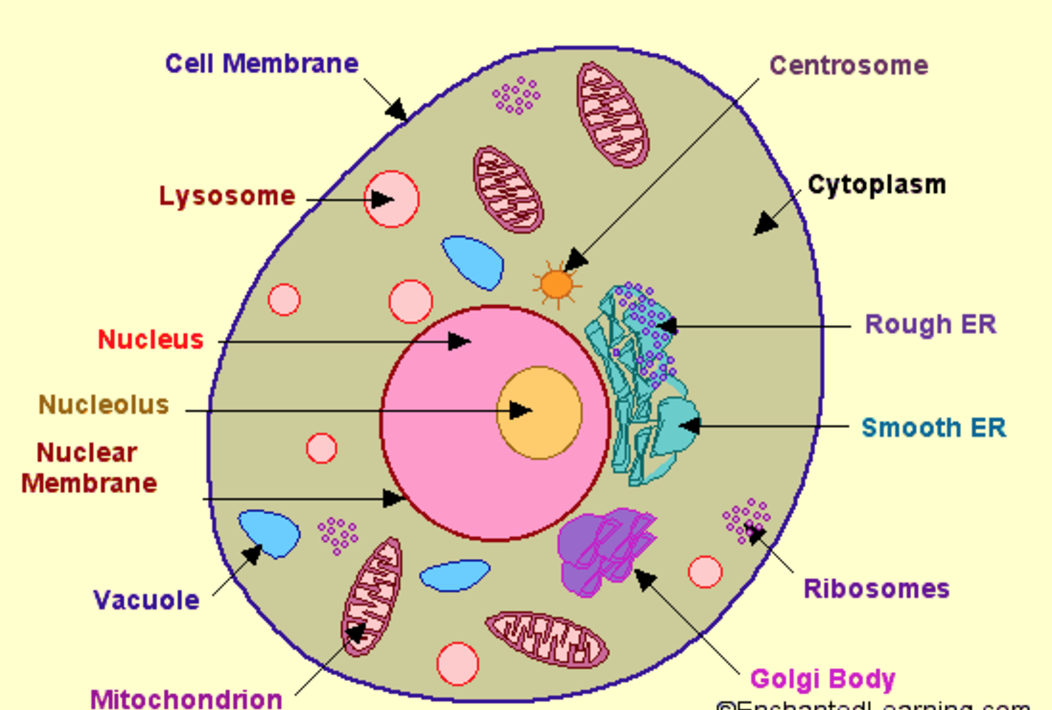
A brief explanation of the different parts of an animal cell along with a well-labelled diagram is mentioned below for reference. Also Read Different between Plant Cell and Animal Cell Well-Labelled Diagram of Animal Cell The Cell Organelles are membrane-bound, present within the cells.

NCERT Class 9 Science Solutions Chapter 5 The Fundamental Unit of Life
Animal Cell: Structure, Parts, Functions, Labeled Diagram June 6, 2023 by Faith Mokobi Edited By: Sagar Aryal An animal cell is a eukaryotic cell that lacks a cell wall, and it is enclosed by the plasma membrane. The cell organelles are enclosed by the plasma membrane including the cell nucleus.
Cell Membrane Images Worksheet Answers
cell, in biology, the basic membrane-bound unit that contains the fundamental molecules of life and of which all living things are composed. A single cell is often a complete organism in itself, such as a bacterium or yeast. Other cells acquire specialized functions as they mature. These cells cooperate with other specialized cells and become.
Education 645 High School Biology
Cells are the fundamental unit of life. All living things are composed of cells. While there are several characteristics that are common to all cells, such as the presence of a cell membrane, cytoplasm, DNA and ribosomes, not all cells are the same. Prokaryotic cells lack a nucleus and membrane-bound organelles.

Labeled Animal Cell Diagram
The plasma (cell) membrane separates the inner environment of a cell from the extracellular fluid. It is composed of a fluid phospholipid bilayer (two layers of phospholipids) as shown in figure 4.1.2 4.1. 2 below, and other molecules. Not many substances can cross the phospholipid bilayer, so it serves to separate the inside of the cell from.

View 20 All Parts Of An Animal Cell Labeled Eporali Wallpaper
1. Plasma membrane: a selective barrier which encloses a cell (plant and bacteria cells also contain a cell wall ). 2. Cytosol: located inside the plasma membrane, this is a jelly-like fluid that supports organelles and other cellular components. 3. Cytoplasm: the cytosol and all the organelles other than the nucleus. 4.
Learn the parts of a cell with diagrams and cell quizzes Kenhub
Cell Parts ID Game. Test your knowledge by identifying the parts of the cell. Choose cell type (s): Animal Plant Fungus Bacterium. Choose difficulty: Beginner Advanced Expert. Choose to display: Part name Clue. Play.
Human cell diagram Etsy
A cell consists of three parts: the cell membrane, the nucleus, and, between the two, the cytoplasm. Within the cytoplasm lie intricate arrangements of fine fibers and hundreds or even thousands of miniscule but distinct structures called organelles. Cell membrane Every cell in the body is enclosed by a cell ( Plasma) membrane.

[DIAGRAM] Diagram Of Cytosol
Figure 6.4 Animal cell mitosis is divided into five stages—prophase, prometaphase, metaphase, anaphase, and telophase—visualized here by light microscopy with fluorescence. Mitosis is usually accompanied by cytokinesis, shown here by a transmission electron microscope. (credit "diagrams": modification of work by Mariana Ruiz Villareal; credit "mitosis micrographs": modification of work by.
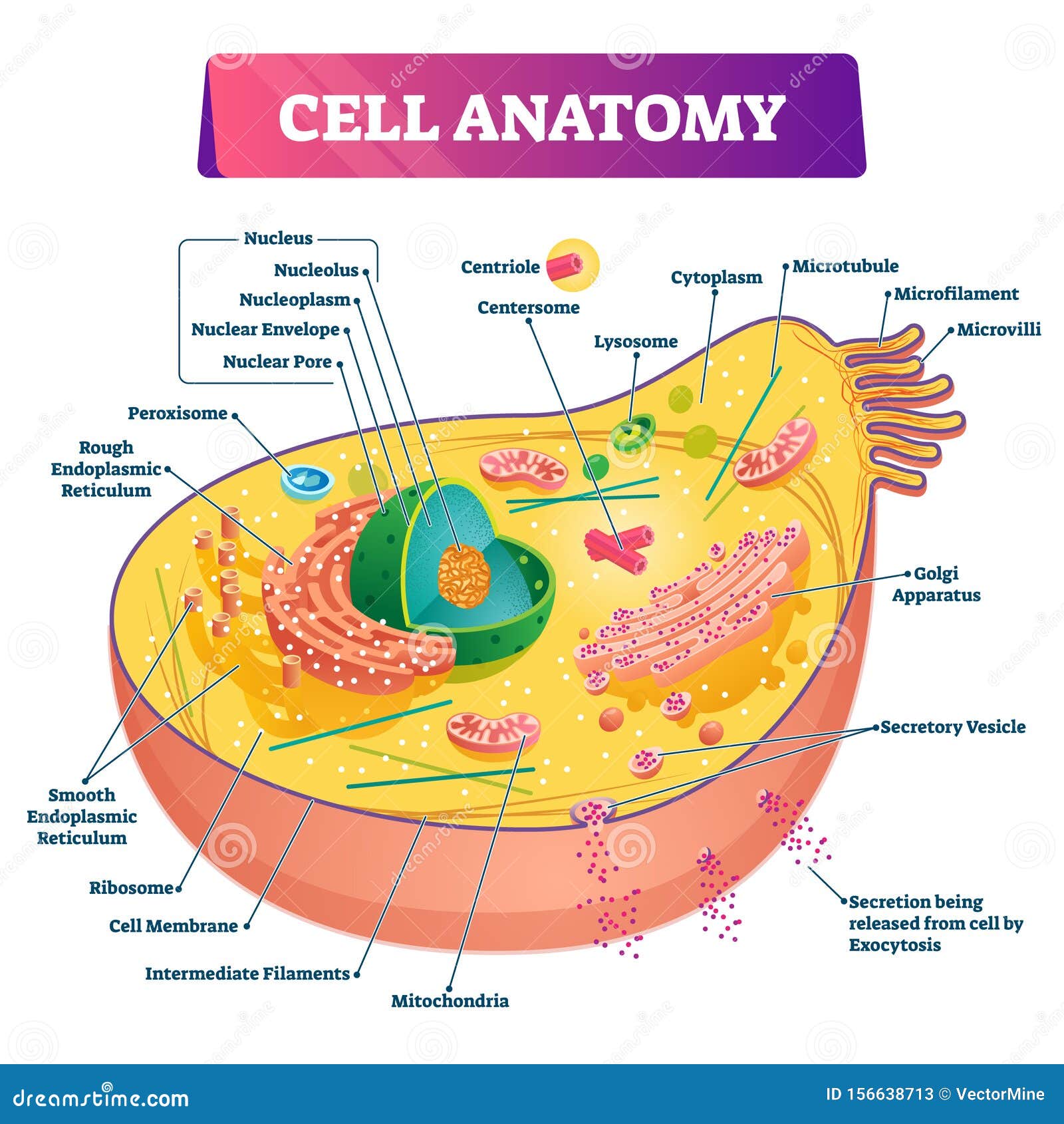
Cell Anatomy Vector Illustration. Labeled Educational Structure Diagram
Middle school biology - NGSS > > Cell parts and functions Cell parts and functions Google Classroom Review your understanding of cell parts and functions in this free article aligned to NGSS standards. Key points: All cells have a cell membrane that separates the inside and the outside of the cell, and controls what goes in and comes out.
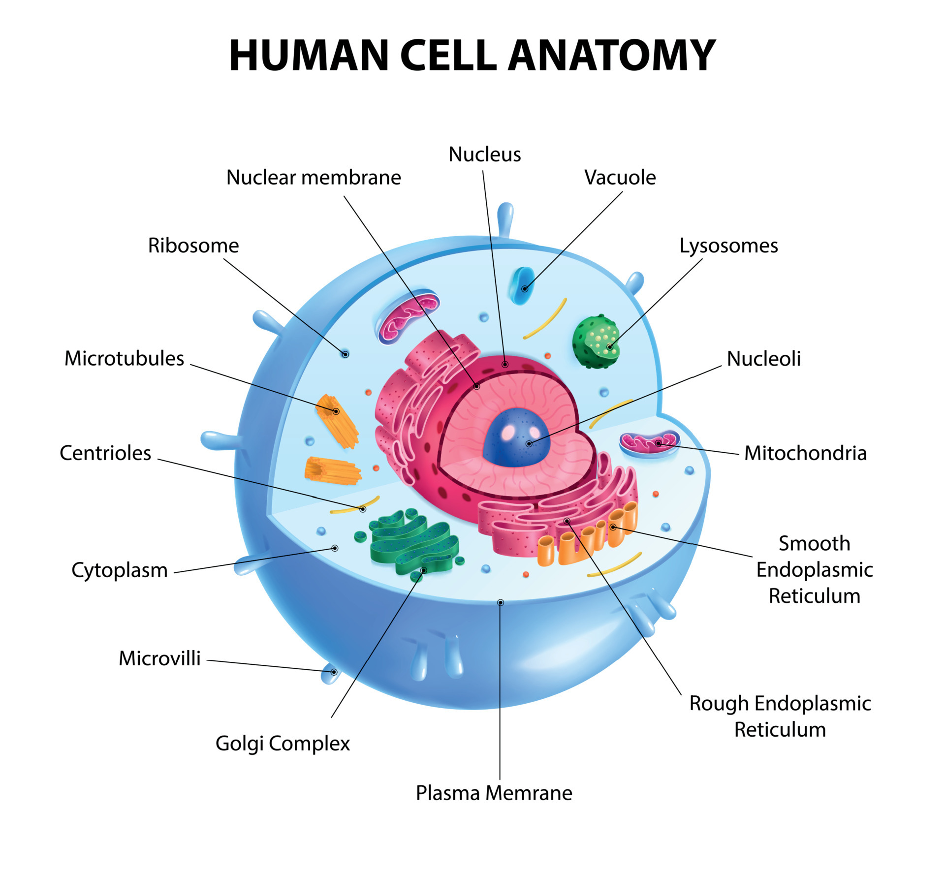
Human Cell Diagram 6406474 Vector Art at Vecteezy
Unit 1 Intro to biology Unit 2 Chemistry of life Unit 3 Water, acids, and bases Unit 4 Properties of carbon Unit 5 Macromolecules Unit 6 Elements of life Unit 7 Energy and enzymes Unit 8 Structure of a cell Unit 9 More about cells Unit 10 Membranes and transport Unit 11 More about membranes Unit 12 Cellular respiration Unit 13 Photosynthesis
